INTRODUCTION: WHERE TO START?
This is one of the most challenging questions when encountering a difficult case. Patients may enter our office hoping to fix an aesthetic problem when their dental issues require far more attention than veneers or crowns alone. Every case truly begins as a “fact finding mission.” We must ask, “How did this patient get into this dental situation?” and evolve into, “Where do we want to go from here?” As dentists, we owe it to our patients to involve the best minds to help us find the way. Together we can work wonders in seemingly impossible cases.
Severe wear cases provide more challenges than most. Are we dealing with abrasion, erosion, attrition, or a combination of factors?1 Will orthodontics be needed? Can we merely treat anterior teeth, or is a full-mouth reconstruction necessary? To create a treatment plan, we must have a diagnostic regimen; an organized step-by-step approach simplifies the process.2
At the heart of a successful diagnostic regimen is a comprehensive examination. This includes an in-depth interview with the patient, an accurate medical history, and an all-encompassing exam including the teeth, periodontium, muscles, and joint. It is essential to recognize the implications of occlusal disharmony before treatment begins, particularly in complex cases.3 Full-mouth radiographs are essential, and in many cases, doppler, CT scan, and/or magnetic resonance imaging are necessary technologies to support the examination, diagnosis, and treatment planning process. Collaboration with specialists and knowledgeable lab technicians makes the treatment more predictable.
The author strongly advises postgraduate training at an advanced learning center (such as the Dawson Academy or Scottsdale Center), as well as staying abreast of new ideas by reading dental journals. Another worthwhile organization, the American Academy of Cosmetic Dentistry and its affiliates, allows one to learn how cosmetic dentistry intersects with comprehensive care.
In a severe wear case, it is essential to use face-bow-mounted diagnostic casts to determine if the patient is occluding in centric relation (CR), and if not, how to get there. An occlusal splint or anterior deprogrammer is an aid in helping determine the causes of the problem and how to correct it. In many cases, patients will suddenly discover that temporomandibular disease (TMD) symptoms disappear that they never knew they had. An aesthetic, comfortable and functionally sound dentition should always be our goal.
Dr. Peter Dawson, founder of the Dawson Academy, has suggested that the goal of treatment of excessive wear should include 6 factors:4
- Equal intensity contacts on all teeth in a verifiable CR
- Anterior guidance in harmony with the patient’s normal functional jaw movements
- Disclusion of all posterior teeth when in protrusive and lateral motion away from CR
- Restoration of any tooth surfaces that have wear through the enamel
- Counseling of the patient to understand that teeth should not be in contact when chewing is not taking place
- Nighttime use of an occlusal splint appliance after therapy is completed.
The sequencing of care involves a plan to systematically reach these goals in an organized way, keeping specialists, the dental laboratory team, and the patient “in the loop” throughout the course of treatment. Stability will be brought about by anterior guidance with posterior disclusion, within the patient’s envelope of function.5
The following report shows how a difficult case can be compartmentalized into a series of steps, leading to a successful result. In addition, it will serve to demonstrate how collaboration with specialists and knowledgeable dental laboratory technicians can pave the way.
CASE REPORT
Diagnosis and Treatment Planning: Consultation Phase
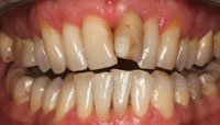
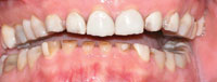
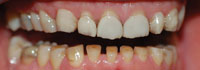
A 37-year-old male patient presented with a desire to replace his 10-year-old maxillary porcelain veneers. He stated that they had become chipped and discolored over time. A visual exam revealed multiple issues. His upper veneers were indeed chipped, stained, and “patched” with composite resin. The clinical crown on all of these veneers was shallow, causing him to squint in order to show his front teeth. His lower incisors showed excessive wear and exhibited only 2.0 to 3.0 mm of tooth structure above the mandibular gingival margins. There was a reverse curve of Spee and there were obvious spaces between mandibular anterior teeth. His lower anteriors were completely obscured by the upper teeth in closure (Before Image and Figures 1 to 3).
 |
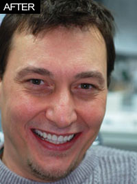 |
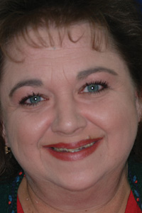
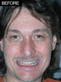
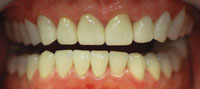
| Before Image. Preoperative photo shows the patient struggling to show upper teeth when smiling. Note chipped and broken 10-year-old veneers and composite resin repairs. | After Image. In full smile, note the pleasing appearance of the upper anteriors and visible lower teeth. The patient reported being very comfortable. As in all severe wear cases, we have emphasized the need to wear a nightguard every night. |
 |
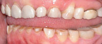 |
| Figure 1. This preoperative view shows the extent of wear on lower anterior teeth as well as gingival heights of these teeth. Virtually all incisal enamel had been worn away. | Figure 2. This view shows a reverse curve of Spee and the flattening of the posterior teeth. Note the apparent abfraction lesion on tooth No. 5. Note also that the upper veneers have chipped and other restorations have broken. |
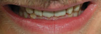 |
| Figure 3. In this view, we can see that the mandibular anterior teeth completely disappear during closure and the patient’s bite is “locked in,”causing a restricted envelope of function. |
 |
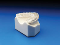 |
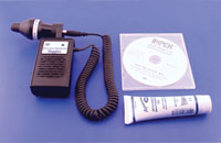
| Figure 4. A temporomandibular joint (TMJ) Doppler (Great Lakes Orthodontics) was used to listen to joint sounds. The patient exhibited crepitus and an occasional click in translation and none in rotation of the joint. (Photo courtesy of Great Lakes Orthodontics.) | Figure 5. The patient wore a full-contact centric relation (CR) splint (Dawson Design) 24 hours a day, removed only for eating and cleaning for 8 weeks. He was surprised at how much better he felt during this period. He had not realized that he had temporomandibular disease symptoms until they disappeared. (Photo courtesy of Great Lakes Orthodontics.) |
An interview with the patient revealed that he had occasional pain in the left and right temporomandibular joint (TMJ) areas. He had discomfort upon palpation of the facial muscles in the left and right superficial masseters and temporalis muscles as well as anterior branches of the sternocleidomastoid. Using a TMJ Doppler (Great Lakes Orthodontics) (Figure 4), crepitus was noticed on both sides with a translational click on the right. It was impossible to establish a definitive CR, and there was pain upon occlusal loading. No teeth showed signs of mobility, and a periodontal exam showed minimal pocketing. The patient agreed to wear a CR bite splint with built-in anterior guidance (Great Lakes Orthodontic Laboratory) (Figure 5) for 2 to 6 months for 24 hours daily (only to be removed during cleaning and meals.) This was planned to better diagnose TMJ issues and to allow us to ultimately mount the case in CR. Initial mounted models and photos were sent to the team at Bayview Dental Laboratory, and I discussed wax-ups and case sequencing personally with Buddy Shaefer, CDT, at the laboratory. Our first goal would be to establish a repeatable CR and to minimize TMD symptoms.
Preliminary Phase
The bite splint was seated and adjusted until all his teeth were in occlusion and posterior disclusion was evident in protrusive and lateral excursions. After several adjustments, the patient was very comfortable with the splint, and the jaw tightness he had grown used to over the years had dissipated, leaving him symptom-free. After 8 weeks of wearing the appliance full time, it was now possible to use bimanual manipulation to determine CR and his envelope of function (Dawson Design). The author has also found that a Lucia Jig (Great Lakes Orthodontics) (Figure 6) or Leaf Gauge (Great Lakes Orthodontics) (Figure 7) are other good tools to assist in finding CR (as suggested by Dr. Frank Spear).6 The patient was finally asymptomatic when loading the joints. A Denar Combi Articulator (Whipmix) and Denar Slidematic Facebow (Whipmix) was used to mount the case in centric relation. It was also noticed that there was a huge slide off his first and second molars that was shifting his bite anteriorly. We decided that removing interferences and creating a bite that would be in line with CR was possible with crown and bridge restorations from teeth Nos. 3 to 15 and 18 to 31. Opening the bite approximately 1.0 mm would help us create a healthy aesthetic occlusion without the need for orthodontics.
 |
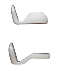 |
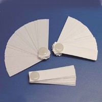
| Figures 6 and 7. The Lucia Jig (Figure 6) and Leaf Gauge (Figure 7) (Great Lakes Orthodontics) provide excellent ways to determine CR in a patient when bimanual manipulation is not possible. In severe wear cases, the author prefers long-term use of a CR splint to allow symptom-free loading of the joint. Another alternative for less complicated bite cases is 3-day use of an anterior deprogrammer appliance and use of the deprogrammer for bite relations instead of the Leaf Gauge or Lucia Jig. (Photos courtesy of Great Lakes Orthodontics.) |
 |
 |
| Figure 8. Following crown-lengthening surgery, more clinical crown was exposed on the lower anterior teeth to allow for better retention for crowns as well as a better cosmetic result. | Figure 9. The final result shows a healthy dentition with normal size lower anterior teeth. Note the more natural gingival heights and pleasing appearance. |
 |
| Figure 10. Note the changes since Figure 3. When the teeth are in contact, the lower teeth are visible. Our goal of equal contacts in CR and disclusion in all excursions has been satisfied. |
When presented with occlusal wear issues, we must determine how many teeth need to be restored to create an occlusion without posterior interferences. Wax-ups are needed in order to determine outside the mouth whether this goal is indeed possible. Altering anterior inclines can help create this situation, but often it is also necessary to change the posterior architecture by equilibration and/or new posterior restorations. In this case, it was deemed essential to restore posterior teeth and remove restorations, which had resulted in the anterior “slide.”
The patient consulted with Dr. Lawrence Tesser (a periodontist) who performed gingival surgery on teeth Nos. 4 to 13 and Nos. 20 to 29 to create more clinical crown length for restorative and cosmetic reasons (Figure 8). Following the crown-lengthening surgery, new models were mounted and sent to the dental laboratory team to do the wax-ups. These would be used to create temporary crowns (Integrity [DENTSPLY Caulk]) after preparing upper and lower teeth on consecutive days. In addition, preparation and reduction guides were made from the wax-ups. After the preparations were completed, the provisional crowns were fabricated (using a putty stent made from an accurate model of the diagnostic wax-up) and cemented with a noneugenol temporary cement (Tempbond NE [Kerr]). Three days later, we adjusted his bite (in CR), including in excursions, and then allowed him time to accommodate.
Impression and Provisional Phase
Three months later, the temporaries and occlusion were still stable. We removed his temporary crowns, packed retraction cord (Ultrapak Cord [Ultradent Products]), and took a vinyl polysiloxane impression of the uppers (Flexitime [Heraeus Kulzer]). Next, a bite registration (Futar D [Kettenbach]) and stick-bite was taken. We created a new lower model of the provisionals using Silgimix (Sultan Healthcare). Then, we again used the Denar Slidematic Facebow (Whipmix) for mounting and sent everything, including photos, to our dental laboratory team. We decided to use porcelain-zirconia restorations due to their strength and cosmetic appeal. New provisionals (Integrity) were made from the putty stent and cemented with a mixture of Tempbond NE and a small amount of Vaseline.
Delivery and Completion
Several weeks later, we removed the provisionals and cemented (Fuji Plus [GC America]) the upper crowns in groups of 4 at a time. We then removed the lower provisionals and took lower full-arch impressions and a new upper model in the same manner as with the upper case.
Two weeks later, the lower crowns were cemented in the same manner as the maxillary crowns. The patient felt comfortable with each set of provisionals and the final crowns (Figures 9 and 10 and After Image) and remains comfortable 2 years later.
CLOSING COMMENTS
As dentists, we face difficult cases every day in our offices. We must determine where we “want to go” with each case, and then discover how we can reach that goal. With the general dentist as the coordinator, a team concept will help us facilitate a well-focused plan. Therefore, it is important to foster a team of professionals who we can trust to help us achieve our goals for the patient. This collaborative effort can create long-term results that we and our patients can be proud of for years to come.
Acknowledgement
The author would like to thank Buddy Shaefer and Walt Richardson (Bayview Dental Laboratory) and my Dawson Academy mentors for teaching the concepts of the comprehensive exam and complete dentistry.
References
- Dudney TE. Treating erosive tooth wear with all-ceramic restorations. Dent Today. 2009;28:100-105.
- Hess LA. Determining the correct vertical and horizontal incisal edge position. Inside Dentistry. 2009;4:26-34.
- Ruiz JL, Coleman TA. Occlusal disease management system: the diagnosis process. Compend Contin Educ Dent. 2008;29:148-156.
- Dawson PE. Evaluation, Diagnosis, and Treatment of Occlusal Problems. 2nd ed. St. Louis, MO: Mosby; 1989.
- Lerner J. A systematic approach to full-mouth reconstruction of the severely worn dentition. Pract Proced Aesthet Dent. 2008;20:81-87.
- Spear F. Centric Relation: Bite Records . Scottsdale, AZ: Spear Education; 2011.
Dr. Auster is president and founder of the Empire State Academy of Cosmetic Dentistry: the New York, New Jersey, and Pennsylvania affiliate of the American Academy of Cosmetic Dentistry (AACD). He graduated in 1980 from the University of Pennsylvania School of Dental Medicine. He is a sustaining member of the AACD and winner of an outstanding service award from the AACD. He is a founding alumnus of the Dawson Academy. Dr. Auster has an aesthetic and restorative practice with 2 partners in Pomona, NY. He can be reached at (845) 364-0400 or via e-mail at drpauster@gmail.com.
Disclosure: Dr. Auster reports no disclosures.