INTRODUCTION
Extractions can be time-consuming and also stressful for the practitioner and the patient. Traditionally, elevators in various sizes and shapes have been utilized to luxate the tooth prior to the use of a forcep to extract the tooth.1-3 These instrument tips are inserted mesially and distally where sufficient alveolus is present that can tolerate the leveraging force applied with the elevator. Unfortunately, when an extraction is carried out with the forceps, the tooth is rotated in a buccal-lingual direction, which is the opposite direction that was applied to luxate the tooth with the elevator.
Periotomes were introduced approximately 25 years ago to improve luxation as an adjunct to use of the elevators.4 These instruments had thinner, more delicate tips intended to be utilized differently then elevators and were designed to be placed into the periodontal ligament (PDL) space with pressure applied in a vertical direction along the root’s long axis.5-7 The tips were provided in 3 orientations to allow access to different areas of the mouth. The periotome was pushed into the PDL, then withdrawn and moved to an adjacent position around the root and pushed apically. This process was repeated while working circumferentially around the root. An elevator could be used next, or, if mobility was noted, a forcep could be used to complete the extraction process. One caution with periotomes: due to the thinner tips on these instruments, if one uses them like an elevator to apply leveraging forces to the root, breakage of the instruments’ tips will occur.
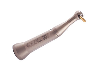 |
| Figure 1. Luxator LX (Directa Dental) power periotome with tip in handpiece. |
More Efficiency with a Power Periotome
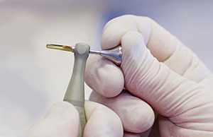
To improve the process, a unique power periotome—the Luxator LX (Directa Dental)—was developed. It fits a standard “E” connector used for dental handpieces to provide activation of the tip (Figure 1) and, unlike hand periotomes with their fixed tips, the Luxator LX tips freely rotate circumferentially. This self-directing tip allows the practitioner to place it into the PDL and, when activated, walk the tip around the tooth without the need to remove it for repositioning (Figure 2). When activated, the tip moves vertically in a reciprocating motion. These tips are not intended for use as elevators. If they are used as elevators, such forces applied will damage not only the Luxator LX periotome tips, but may also damage the alveolar bone at the buccal or lingual crest.
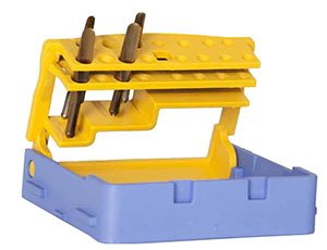
The Luxator LX periotome tips have a concave inner and convex outer surface in cross section, available in 2.0-mm and 3.0-mm wide versions and in short and long lengths (Figure 3). The tips are very thin and sharp, and, when inserted into the PDL space, they will separate the ligaments between the root and alveolus while compressing the alveolar bone of the socket to create space and mobilize the tooth. The titanium-nitride coating makes the tips more durable. The hardening process also gives excellent long-term durability to the sharpness of the tip. The tips are provided in an autoclavable carrier to prevent losing them between uses on patients (Figure 4). To remove the tip from the handpiece, a metal plunger is provided. Insert the plunger into the back of the handpiece head to pop the tip out (Figure 5).
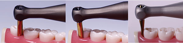 |
| Figure 2. The tip is placed into the periodontal ligament (PDL) space of the tooth to be extracted, activated via the foot rheostat, then slowly walked around the tooth while applying light apical pressure. |
The recommended speed when operating the Luxator LX is 4,000 rpm, and the author recommends starting at a low speed and increasing if the tip is not advancing into the PDL with light apical pressure. Speeds faster than 4,000 rpm will not make extractions easier or faster, but may cause uncomfortable vibrations for the patient. Higher speeds may also damage the Luxator LX contra-angle handpiece.
CLINICAL CASE EXAMPLES
Extraction of Intact Teeth
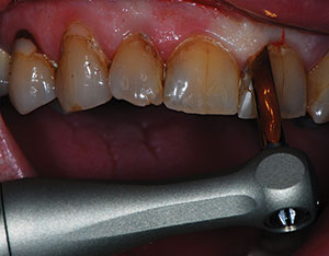
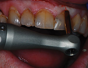
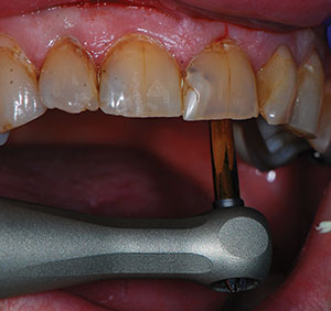
When an intact tooth is to be extracted, the appropriate Luxator LX periotome tip is selected and inserted into the handpiece. The tip is placed into the PDL space at the mesial-interproximal-buccal aspect and light apical pressure is exerted (Figure 6). The tip is maintained in a position that aligns it with the long axis of the tooth’s root. With the tip in the PDL space, the rheostat is then activated, and the tip is walked to the distal-interproximal-buccal aspect while maintaining light apical pressure (Figure 7). The handpiece is deactivated, and the tip is then moved to the lingual and reinserted into the PDL space. Next, the handpiece is reactivated and the tip is moved to toward the interproximal area where the process initially started (Figure 8). Mobility in the buccal-lingual direction is checked and, if sufficient, an extraction forceps is then utilized to complete the extraction process. If the tooth is not sufficiently mobile, the tip is reinserted into the PDL space and the process is simply repeated.
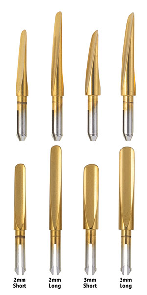 |
| Figure 3. Luxator LX periotome tips, shown here at 2 angles to emphasize how the concave shape of the tips match the contours of the root(s). |
 |
 |
| Figure 4. The auotclavable Luxator LX tip holder. | Figure 5. The Luxator plunger being used to remove the periotome tip from the handpiece. |
Extraction of Roots and Nonintact Teeth
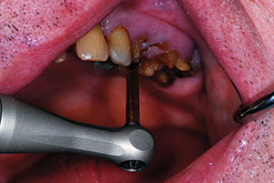
The Luxator LX periotome can be utilized on nonintact teeth and residual roots with a flapless or flap procedure. The technique is identical to the one used on intact teeth. However, without the coronal tooth structure present, the tip can be moved circumferentially, without having to remove and reposition the tip around the proximal contact with the adjacent tooth. The tip is introduced on the buccal (Figure 9) and, once activated, it is moved circumferentially around to the lingual (Figure 10) to complete the process. An elevator (such as the Luxator Forte Elevator [Directa Dental], Luxating Elevator [Hu-Friedy], or Zators [Zoll-Dental]) or a similar instrument can be applied into the widened PDL space to further luxate the remaining root before moving to a forceps or rongeur to complete the extraction process.
 |
 |
| Figure 6. In this clinical case, the tip was introduced into the mesial buccal of the intact central incisor that was to be extracted, then activated. | Figure 7. While activated, the periotome tip is walked slowly to the distal buccal while applying light apical pressure. |
 |
 |
| Figure 8. The periotome tip was reoriented to the palatal and inserted into the PDL space at the distal palatal, then activated and walked to the mesial palatal. | Figure 9. In this clinical case, the Luxator LX periotome tip was inserted into the PDL space at the buccal of the root to be extracted and activated. |
 |
| Figure 10. The Luxator LX periotome tip was walked around the root to be extracted while activated, applying light apical pressure to luxate the tooth. |
CLOSING COMMENTS
Tooth extractions can be a stressful appointment for both the patient and the practitioner. When the practitioner rushes the process, the tooth being extracted may fracture, thus complicating the procedure and requiring more time and effort to retrieve any remaining root in the alveolus. The key to tooth extraction is taking time to mobilize the root so that it can be removed as atraumatically as possible. The Luxator LX power periotome is an improvement over hand periotomes, as it requires less hand force by the practitioner to luxate the root and is more efficient in motion. Additionally, it provides a gentler experience for the patient, helping to alleviate fears that may have caused them to be hesitant with future dental treatments.
References
- Malden N. Surgical forceps techniques. Dent Update. 2001;28:41-44.
- Krekmanov L. Extraction with an elevator [in Swedish]. Tandlakartidningen. 1974;66:1365-1370.
- Fries R, Platz H. Use of the elevator in tooth extractions [in German]. Osterr Z Stomatol. 1974;71:334-339.
- Thomson PJ. Minimising trauma in dental extractions: the use of the periotome. Br Dent J. 1992;172:179.
- Sharma SD, Vidya B, Alexander M, et al. Periotome as an aid to atraumatic extraction: a comparative double blind randomized controlled trial. J Maxillofac Oral Surg. 2015;14:611-615.
- Kaijin H, Yongfeng L. Application of micro-power system in the surgery of tooth extraction [in Chinese]. Hua Xi Kou Qiang Yi Xue Za Zhi. 2015;33:1-5.
- Levitt D. Atraumatic extraction and root retrieval using the periotome: a precursor to immediate placement of dental implants. Dent Today. 2001;20:53-57.
Dr. Kurtzman is in private general practice in Silver Spring, Md, and is a former assistant clinical professor at the University of Maryland and a former assistant program director for the American Academy of Implant Dentistry implant maxi-course at Howard University College of Dentistry (Washington, DC). He has earned Fellowships in the AGD, American Academy of Implant Prosthodontics, American College of Dentists, International Congress of Oral Implantologists (ICOI), Pierre Fauchard, and the Academy of Dentistry International; Masterships in the AGD and ICOI; and Diplomate status in the ICOI and American Dental Implant Association. He has lectured internationally on the topics of restorative dentistry, endodontics, implant surgery, removable and fixed prosthetics, and periodontics and has more than 525 published articles globally. He has been honored to be included in the Leaders in Continuing Education directory in Dentistry Today annually since 2006 and was featured on its June 2012 cover. He can be reached via email at dr_kurtzman@maryland-implants.com.
Disclosure: Dr. Kurtzman reports no disclosures.
Related Articles
6-Part Series Explores the Evolution of Comprehensive Care
Laser Endodontic Debridement and Canal Disinfection
Laser Troughing to Improve Scanning and Impressions